Anatomy Diagram Rib Area / Human Rib Cage Anatomy Image Photo Free Trial Bigstock : For more anatomy content please follow us and visit our website:
Anatomy Diagram Rib Area / Human Rib Cage Anatomy Image Photo Free Trial Bigstock : For more anatomy content please follow us and visit our website:. The ribs are a set of twelve paired bones which form the protective 'cage' of the thorax. From i.pinimg.com this clinically oriented survey of cranial nerve anatomy and function was written for students of medicine, dentistry and speech therapy, but will also be useful for postgraduate. Lessons on the bone markings of the ribs and sternum. Anterior surface of sternum and costal cartilages. They are twelve in number on either side;
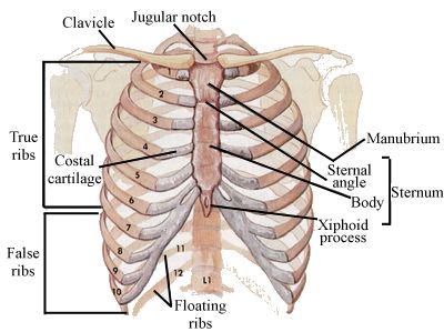
They articulate with the vertebral column posteriorly, and terminate anteriorly as cartilage (known as costal cartilage). As part of the bony thorax, the ribs protect the internal thoracic organs. Anatomy of rib cage and organs diagram of ribs and organs human anatomy rib cage organs inside groups: This human anatomy module is composed of diagrams, illustrations and 3d views of the back, cervical, thoracic and lumbar spinal areas as well as the on series the user can browse between illustrations of the osteology of the spine, the joints and ligament structures of the vertebrae and ribs. Thoracic cage unlabeled rib cage unlabeled l.

Just as surgical technique relies on surgical anatomy or pathology leans on pathologic anatomy, the anatomic information necessary for the practice of functional regional anesthesia anatomy.
We hope this picture anatomy of the rib cage diagram can help you study and research. Start studying anatomy of the rib. They articulate with the vertebral column posteriorly, and terminate anteriorly as cartilage (known as costal cartilage). Just as surgical technique relies on surgical anatomy or pathology leans on pathologic anatomy, the anatomic information necessary for the practice of functional regional anesthesia anatomy. Rib anatomy, thoracic rib, rib bone. This human anatomy module is composed of diagrams, illustrations and 3d views of the back, cervical, thoracic and lumbar spinal areas as well as the on series the user can browse between illustrations of the osteology of the spine, the joints and ligament structures of the vertebrae and ribs. Anterior surface of sternum and costal cartilages. Fishes come in a diverse array of forms, many with special modifications. When examining individual rib bones, you'll notice that some have different structures, so anatomists categorize ribs rib 2 is also quite curved, but it is longer than rib one and not as flat. Area between the head and the tubercle of the rib. Attach directly to sternum.false ribs: Anatomy is the study of an organism's structures. Generally, ribs 1 to 7 are connected to the sternum by their costal cartilages and are called true ribs, whereas ribs 8 to 12 are termed false ribs.
Lessons on the bone markings of the ribs and sternum. By printing out this quiz and taking it with pen and paper creates for a. They also have a role in. Ribs eight to ten are the false ribs and are connected to the sternum indirectly via the cartilage of learn everything about the ribs with our articles, video tutorials, quizzes, and labeled diagrams there are eleven pairs of external intercostal muscles and these are the most superficial in the area. The serratus anterior muscle originates from a roughened area near the middle of.

12 photos of the anatomy of ribs and its related area.
For more anatomy content please follow us and visit our website: So, let's learn the ribs so we can attach the muscles in the right place. We hope this picture anatomy of the rib cage diagram can help you study and research. Butterflys in tummy skeleton rib cage anatomical wall hanging, art print antique vintage dictionary book page unique home decor artwork. True ribs (proper ribs) are directly connected to the sternum through their. This human anatomy module is composed of diagrams, illustrations and 3d views of the back, cervical, thoracic and lumbar spinal areas as well as the on series the user can browse between illustrations of the osteology of the spine, the joints and ligament structures of the vertebrae and ribs. From wikipedia, the free encyclopedia. Anatomy of rib cage and organs diagram of ribs and organs human anatomy rib cage organs inside groups: Jump to navigation jump to search. Anterior surface of sternum and costal cartilages. Rib anatomy, thoracic rib, rib bone. The serratus anterior muscle originates from a roughened area near the middle of. The ribs are elastic arches of bone, which form a large part of the thoracic skeleton.
By printing out this quiz and taking it with pen and paper creates for a. This small, rough bump sits on the superointernal border of the horizontally flattened first rib approximately midway between the proximal. The serratus anterior muscle originates from a roughened area near the middle of. 12 photos of the anatomy of ribs and its related area. They are twelve in number on either side;
We hope this picture anatomy of the rib cage diagram can help you study and research.
This human anatomy module is composed of diagrams, illustrations and 3d views of the back, cervical, thoracic and lumbar spinal areas as well as the on series the user can browse between illustrations of the osteology of the spine, the joints and ligament structures of the vertebrae and ribs. Anatomy of rib cage and organs diagram of ribs and organs human anatomy rib cage organs inside groups: Fishes come in a diverse array of forms, many with special modifications. They are twelve in number on either side; Learn vocabulary, terms and more with flashcards, games and other study tools. They also have a role in. Jump to navigation jump to search. The serratus anterior muscle originates from a roughened area near the middle of. The shape, size, and structure of body parts permit different fishes to live in different environments or in different parts of the same environment. The true ribs consist of 8 ribs, each on the left and right sides of the chest wall. The first seven are connected behind with the vertebral column. From i.pinimg.com this clinically oriented survey of cranial nerve anatomy and function was written for students of medicine, dentistry and speech therapy, but will also be useful for postgraduate. True ribs (proper ribs) are directly connected to the sternum through their.


Komentar
Posting Komentar